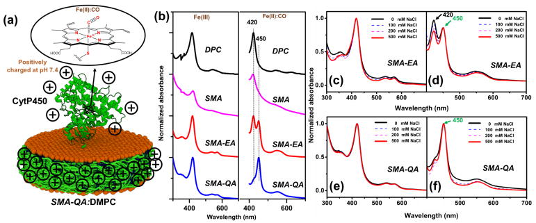
Figure 2. Reconstitution of CytP450 2B4 in polymer nanodiscs.
(a) Schematic showing CytP450 reconstited in a SMA-QA:DMPC nanodiosc. Heme corination sphere of CO bound state. (b) UV-vis absorption spectra of CytP450 reconstituted in different nanodiscs in its ferric state (left column) and ferrous -carbon monoxide complex (right column). UV-vis absorption spectra of cytP450 reconstituted in SMA-EA nanodiscs: (c) in the presence of indicated NaCl concentrations and (d) ferrous carbon monoxide complex (d). UV-vis spectra of cytP450 reconstituted in SMA-QA nanodiscs and (e) in the presence of NaCl, (f) ferrous carbon monoxide complex.

