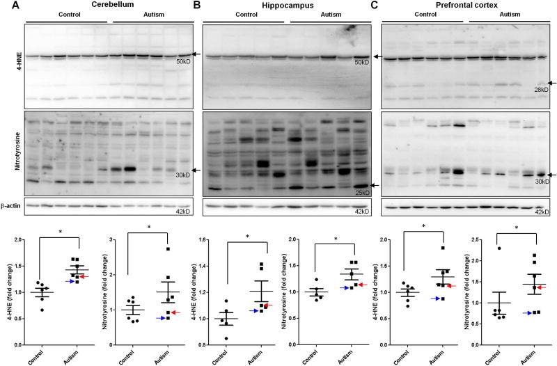
Figure 9.
Lipid peroxidation and nitrotyrosine in the autistic brain. Immunoblot analysis of 4-HNE and nitrotyrosine in the autistic: (A) cerebellum (n=6), (B) hippocampus (n=5) and (C) prefrontal cortex (n=6). Representative images of the immunoblots are shown in top panel. The modified blots were indicated with arrow. The blue arrow indicates sample #4849 and the red arrow indicates sample # 5565. The quantification of immunoblots normalized to β-actin is shown by bar graphs. Values are the means ± SEM. * indicates a significant difference in autistic brains compared to controls (P < 0.05).

