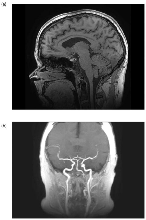
Figure 8.
(a) Sagittal 3D MP-RAGE acquisition with 0.7 × 0.7 × 0.7 mm3 spatial resolution covering the entire brain in 8:25 with the 32-channel brain coil. Other acquisition parameters were: FOV = 22 cm, TE/TR = 3.1/7.3 ms; TI=1000 ms. Image detail of the brain and cerebellum is exquisite. Signal remains uniform and robust down to the level of the C2/C3 cervical interspace. There are only minimal susceptibility artifacts at the interface of the brain and paranasal sinuses (b) Maximum intensity projection of the 3D MP-RAGE acquisition showing high signal within the internal and external carotid arteries, and the proximal vertebral arteries. This bright blood extends more superiorly on the C3T MP-RAGE images, as the IR pulse does not extend into the lower neck and body as it would with conventional whole-body coil excitation.

