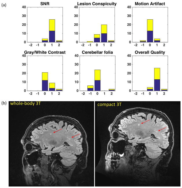
Figure 9.
(a) Results of the right-side Wilcoxon Sign-Rank test from the assessment of 3D CUBE T2-weighted FLAIR image quality by 2 blinded neuroradiologists. (b) Comparison images in a patient with mild cognitive impairment showing white-matter lesions (arrows). Improved SNR and lesion conspicuity with the C3T system is evident from the shorter echo-spacing, resulting in reduced T2 blurring of the FSE echo train and overall improvement in image sharpness. The 8-channel brain coil was used in all instances with TR=7600 ms; 256 × 256 acquisition matrix in a 24-cm FOV; 152 1.2-mm partitions (0.94 mm × 0.94mm × 1.2 mm voxels); TE/TI=93.0 ms/2025 ms for the whole-body system, and TE/TI = 91.3 ms/2060 ms for the C3T system; echo-train length (ETL) = 200; in-plane acceleration R=2. The echo-spacing for the whole-body system was 4824 μs, and 3544 μs for the C3T scanner.

