Fig. 5.
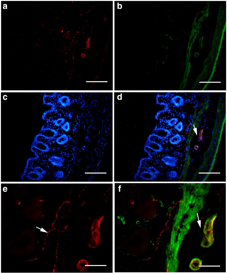
Expression of P2X1 receptors and SPMA in the mouse distal colon. a P2X1 receptor-ir. b SPMA-ir in the same region of a. c DAPI staining in the same region of a. d The merged image of a–c. e A high magnification of the region indicated by an arrow in d, a white arrow indicates a nerve fiber with P2X1 receptor-ir. f The double-labeled image of P2X1 receptor-ir and SPMA-ir in the same region of e, an arrow indicates a double-labeled blood vessel. The scale bars in a–d = 120 μm, in e, f = 60 μm

