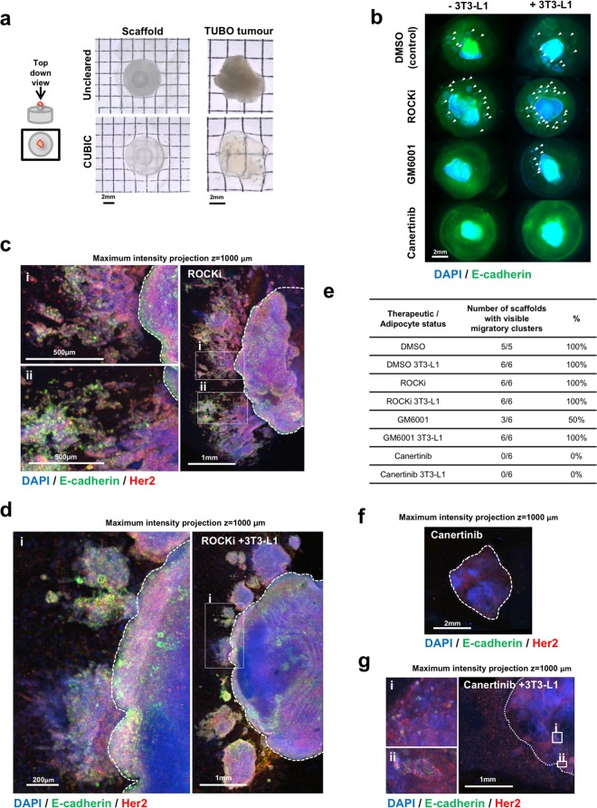
Figure 7.
Optical clearing of TUBO tumours in anisotropic collagen scaffolds and cancer therapeutic testing. (a) Transmission stereoscopic images of uncleared and optically cleared (CUBIC) anisotropic collagen scaffold and TUBO (Her2-neu overexpressing) tumour from a top down view. (b) Fluorescent stereoscopic images of TUBO tumour fragments in scaffolds, cultured for 10 days with/without adipocytes (3T3-L1), treated with DMSO (control), ROCKi (Y-27632), GM6001 or Canertinib, CUBIC cleared and immunostained for E-cadherin (green) and Her2 (not shown). Cell nuclei are stained with DAPI (blue). Clusters of cancer cells that have migrated away from the central tumour fragment are marked with arrowheads. (c,d) Large tile scan z-stack (1 mm depth) confocal microscopy images of ROCKi treated TUBO tumour fragments in anisotropic collagen scaffolds without/with adipocytes (3T3-L1), cleared and stained as described in (b) with DAPI (blue), E-cadherin (green) and Her2 (red). (i) and (ii) show magnified images of migratory clusters of Her2 and E-cadherin positive cells. (e) Quantification of the number of tumour/scaffolds that contain one or more visible migratory cell clusters. (f,g) Large tile scan z-stack (1 mm depth) confocal microscopy images of Canertinib treated TUBO tumour fragments in anisotropic collagen scaffolds without/with adipocytes (3T3-L1), cleared and stained as described in (b) with DAPI (blue), E-cadherin (green) and Her2 (red). (i) and (ii) show few non-migratory E-cadherin and Her2 cells seen in the seeded tumour fragment. No migratory cells were observed in the scaffold with Canertinib treatment at this magnification.

