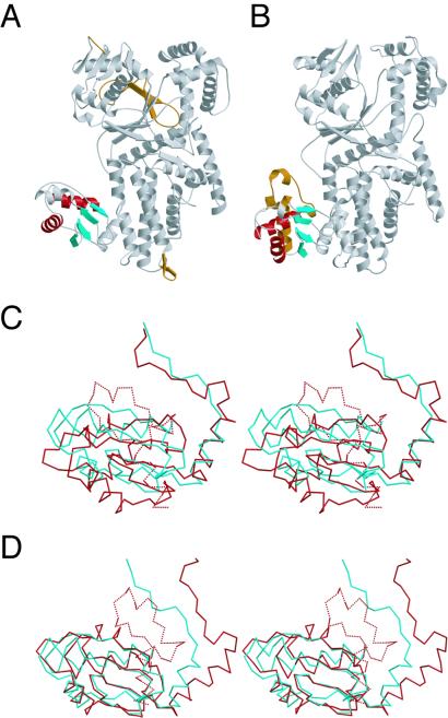
Figure 2.
(A and B) Ribbon diagrams of T. thermophilus ArgRS (A) and S. cerevisiae ArgRS (B). The common antiparallel β-sheets with the lining α helices of the N-terminal domain of the T. thermophilus and S. cerevisiae ArgRSs are colored red for α-helices and cyan for β-sheets, respectively. The additional N-terminal extension of the S. cerevisiae ArgRS and the specific insertions of the T. thermophilus ArgRS are colored brown. (C) Different orientations of the antiparallel β-sheet with the three lining α helices of the N-terminal domain between the T. thermophilus ArgRS (cyan) and the S. cerevisiae ArgRS (red), with the rest of the molecules superimposed (stereo view). The N-terminal extension specific to the S. cerevisiae ArgRS is indicated as dashed lines. (D) Superposition of the antiparallel β-sheet of the N-terminal domain between the T. thermophilus ArgRS (cyan) and the S. cerevisiae ArgRS (red; stereo view).

