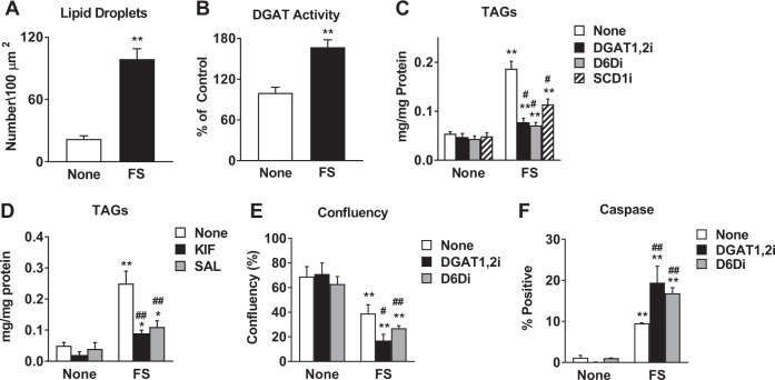
Fig. 6. Accumulation of PUFA-TAGs plays a protective role.
a Nile Red staining was performed on MCF-7 cells treated with 5 μM FS for 48 h. The number of lipid droplets per 100 µm2 were quantified by ImageJ FIJI. b DGAT activity was measured in untreated and FS-treated MCF-7 cells, as described in Materials and Methods section. c TAGs were measured using Triglyceride Colorimetric Assay Kit in MCF-7 cells treated with FS in the presence or absence of DGAT inhibitors (DGAT1,2i, 10 μM each), Δ6 desaturase inhibitor (DGDi, 50 μM), or SCD inhibitor (SCDi, 50 μM). d TAGs were measured in untreated or FS-treated MCF-7 cells in the presence or absence of EnRS inhibitors (KIF, 1 μM or SAL, 50 μM). e, f Confluency and caspase activity were measured following treatment with FS in the presence or absence of DGAT1,2i or D6Di. * P < 0.05 vs none; ** P < 0.01 vs none. # P < 0.05 vs FS alone; ## P < 0.01 vs FS alone

