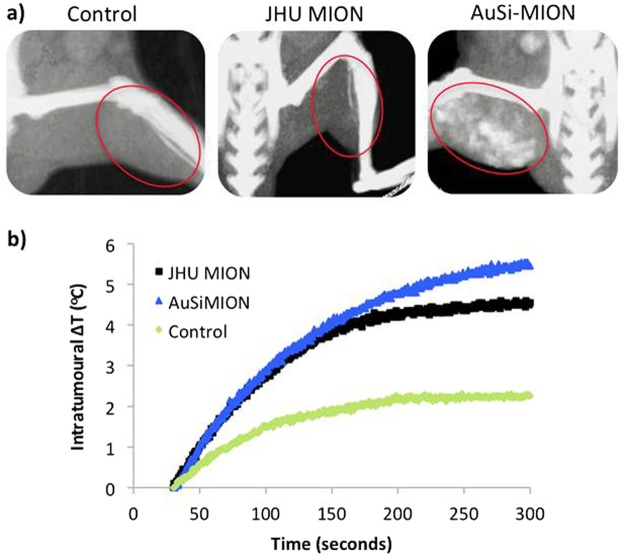
Figure 7.
In vivo imaging and heating. (a) In vivo CT imaging in nude male mice bearing human prostate (LAPC-4) cancer xenograft tumors. Control: saline only injection (left) - the red oval denotes the location of the tumor, which is invisible without added contrast. JHU MIONs (middle): injection concentration of 5.5 mg Fe/cm3 tumor. Iron oxide demonstrates insufficient x-ray contrast with CT rendering the tumor invisible. AuSi-MIONs (right): injection concentration of 5.5 mg Fe/cm3 tumor. The AuSi-MIONs are visible in the tumor indicated by increased signal. (b) Following CT imaging, mice were placed in an AMF device (150 kHz, 40 kA/m) and a ~6°C rise of tumor temperature was measured with RF-resistant optical fiber temperature probe inserted into tumors loaded with either JHU MIONs or AuSi-MIONs.

