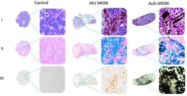
Figure 8.
Histology of prostate tumor xenografts. (a) Mice were euthanized and tumor tissues were collected for staining 72 h post AMF exposure (Row I: H&E, row II: Prussian blue and row III: silver enhancement stain). The control shows no iron oxide or gold present. Tissues from the mouse injected with JHU MIONs show iron oxide particles in the H&E stain, iron staining (blue) with Prussian blue and no response to the silver enhancement stain. Tissues from the mouse injected with AuSi-MIONs show a dark purple color from the gold nanoparticles in the H&E stain and iron staining (blue) with Prussian blue. Dark black staining of the AuSi-MIONs by the silver enhancement stain, which only stains metallic gold or silver, can be seen in row III. Whole tumor images are composites created from separate 4x images; magnified images were obtained at 20x.

