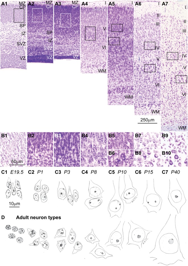
FIGURE 6.
Cytodifferentiation of cortical neurons during development and in adulthood. (A1–A7) Nissl staining of area HL of the rat cerebral cortex during different embryonic (A1: E19.5) and postnatal (A2: P1; A3: P3; A4: P8; A5: P10; A6: P15; A7: P40) developmental stages show the expansion of the cortical plate (CP) and the formation of a eulaminate 6-layered cortex with the addition of new neurons, the progressive enlargement of their cytoplasm and nucleus, and the expansion of the neuropil. (B1–B10) High magnification of insets in (A1–A7) in the middle/deep cortical layers that show parallel changes in chromatin distribution patterns and size of the nucleus and cytoplasm associated with neuron maturation. Differentiation of neurons progresses from deep to superficial layers. (C1–C7) Sketches of developing layer V pyramidal neurons of the rat cerebral cortex from levels shown in (B1–B4,B6,B8,B10). (D) Sketches of neuron types in the adult rhesus macaque; the left panel below (C1) depicts cerebellar granule cells; the panels below (C2–C7) depict progressively larger neurons of the cerebral cortex. In line with what LaVelle (1956), LaVelle and LaVelle (1970) suggested it appears that each stage in the development of nuclear architecture of the largest neurons (series in C) has a structural counterpart in the adult (series in D), in the form of some small neuron that never progressed beyond that stage of maturity. CP, cortical plate; IZ, intermediate zone; MZ, marginal zone; SP, subplate; SVZ, subventricular zone; VL, ventricular zone; WM, white matter. Roman numerals indicate cortical layers. Calibration bar in (A6) applies to (A1–A7). Calibration bar in (B1) applies to (B1–B10). Calibration bar in (C1) applies to (C1–C7) and (D). (This figure is a reexamination of material from a gift of Dr. Alan Peters).

