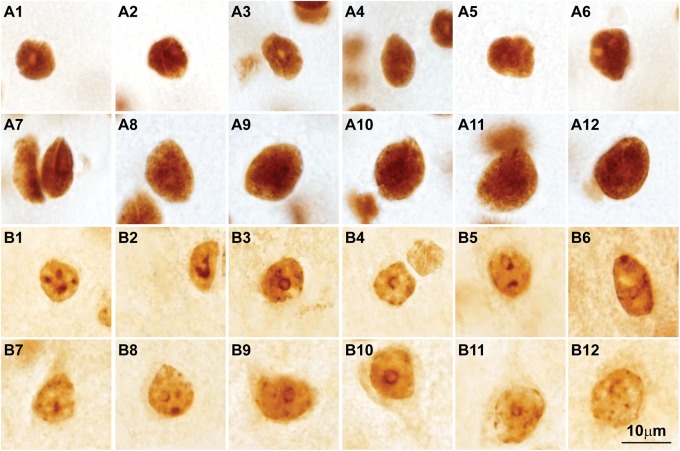
FIGURE 7.
Post-translational histone modifications in the nuclei of cortical neurons. (A1–A12) Photomicrographs of cortical neurons in the primary visual cortex of the rhesus macaque labeled for H3K4met1-2-3 [Anti-mono/di/trimethyl-Histone H3 (Lys4), cat. #04-791, Millipore, Temecula, CA, United States], which shows a modification that facilitates gene expression; the nuclei of small and large neurons show intense H3K4met1-2-3 labeling with empty pockets that correspond to nucleoli and thick heterochromatin clumps. (B1–B12) Micrographs of cortical neurons in the primary visual cortex of the rhesus macaque labeled for H3K9met3 [Anti-trimethyl-Histone H3 (Lys9), cat. #07-442, Millipore], a repressive histone mark for gene expression; the labeling of H3K9met3 highlights heterochromatin grains and clumps with a distribution comparable to Nissl stained neurons (compare with Figures 2B1–B15). Calibration bar in (B12) applies to all panels.

