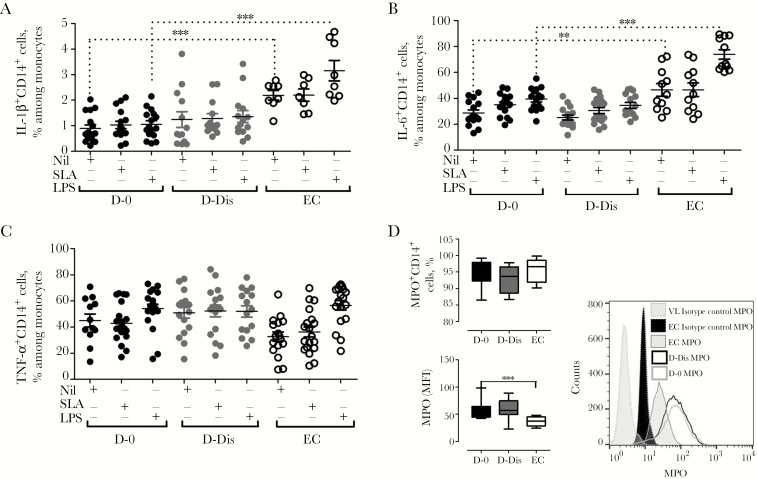
Figure 4.
Analyses of intracellular production of inflammatory cytokines and myeloperoxidase (MPO) during visceral leishmaniasis (VL). A–C, Soluble Leishmania antigen (SLA)– and lipopolysaccharide (LPS)–induced production of interleukin 1β (IL-1β), interleukin 6 (IL-6), and tumor necrosis factor α (TNF-α) by CD14+ cells in cultures of peripheral blood mononuclear cells from patients with VL and healthy endemic controls (ECs) was measured to assess the cytokine-producing ability of monocytes. Analysis was performed on CD14+ cells, and the frequency of CD14+ cells with a particular phenotype was expressed as a percentage of the total number of CD14+ cells. Representative plots indicate the percentage of CD14+ cells expressing IL-1β (A), IL-6 (B), and TNF-α (C) in samples from patients with VL before treatment (D-0; solid black circles) and after 28 days of treatment and prior to discharge (D-Dis; solid gray circles) and in samples from ECs (clear black circles) following stimulation with SLA and LPS. Graphs depict data from 16 paired specimens from the VL group and 11 specimens from the EC group (A and B) and 21 specimens from D-0, 17 from D-Dis, and 20 from the EC group (C). D, Ex vivo intracellular whole-blood staining for MPO showed no changes in the frequency of MPO-expressing CD14+ cells but increased expression of MPO on monocytes in 8 paired specimens from patients with VL, compared with specimens from 10 ECs. Representative histograms displaying MPO mean fluorescence intensities in 1 sample from D-0, 1 sample from D-Dis, and 1 EC sample are shown.

