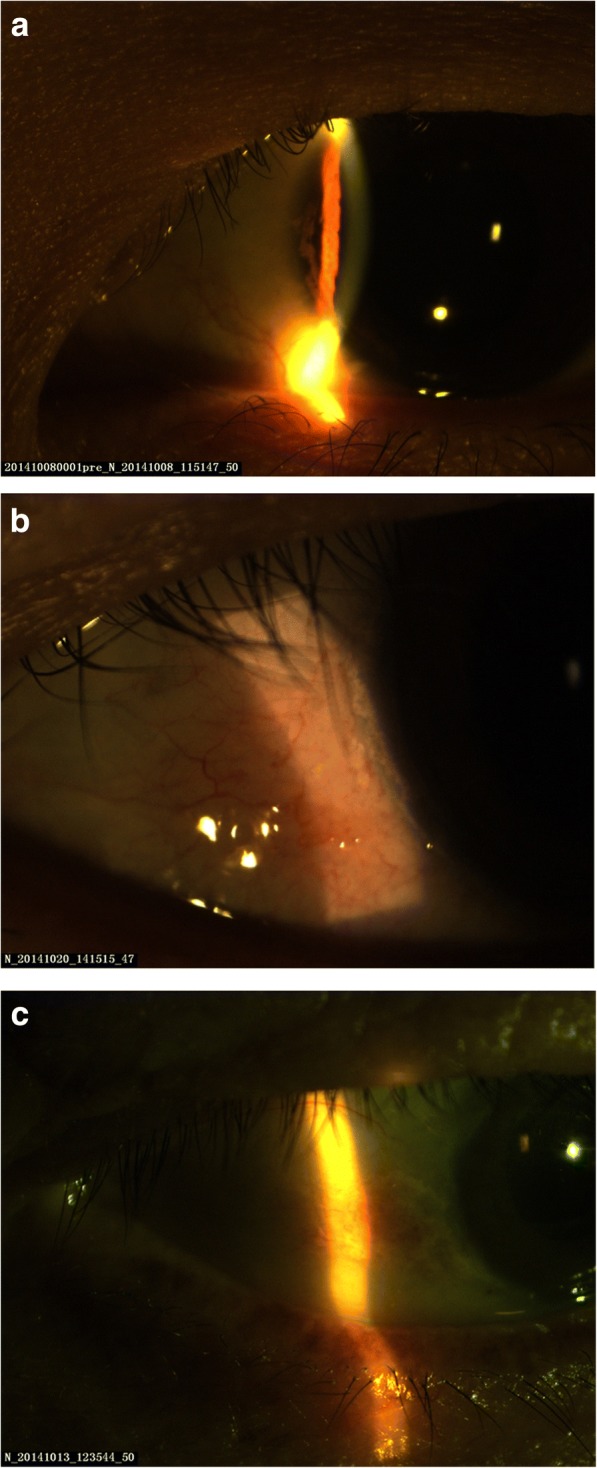
Fig. 1.

Conjunctival and corneal calcification. a In patient A, who had a 7-year HD history, a gray, line-shaped calcium deposit could be seen on the cornea. b In patient B, who had a 3-year HD history, white dot- and line-shaped calcium deposits were observed at the limbus. c In patient C, who had a 6-year HD history, white, block-shaped conjunctival calcium deposits were observed
