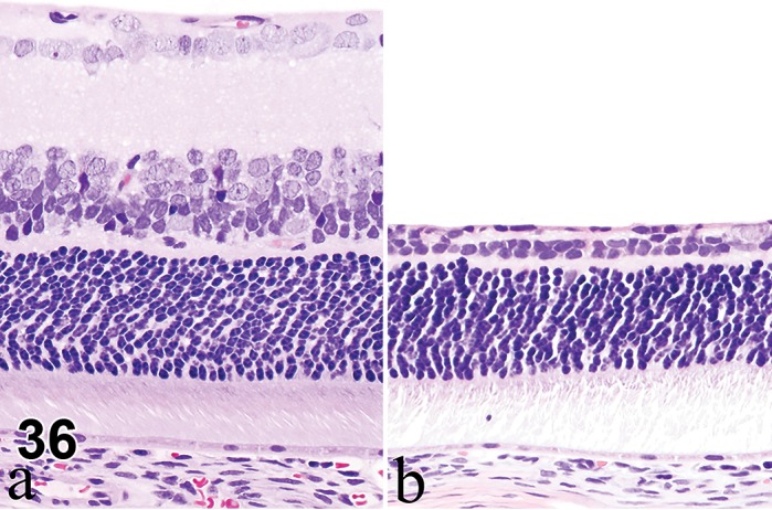
Figure 36.

Atrophy, inner retina; rat. Normal retinal anatomy of control rat (a), compared to retina with atrophy (b). Diffuse atrophy of nerve fiber, ganglion cell, inner plexiform and inner nuclear layers is depicted. Note sparing of outer nuclear layer.
