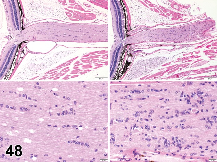
Figure 48.

Increased numbers (“gliosis”), glial cell; optic nerve; mouse. The top and bottom (higher magnification) images on the right depict an increase in numbers of glial cell nuclei dispersed throughout the optic nerve neuropil. Compare to normal optic nerve shown in the top and bottom images to the left.
