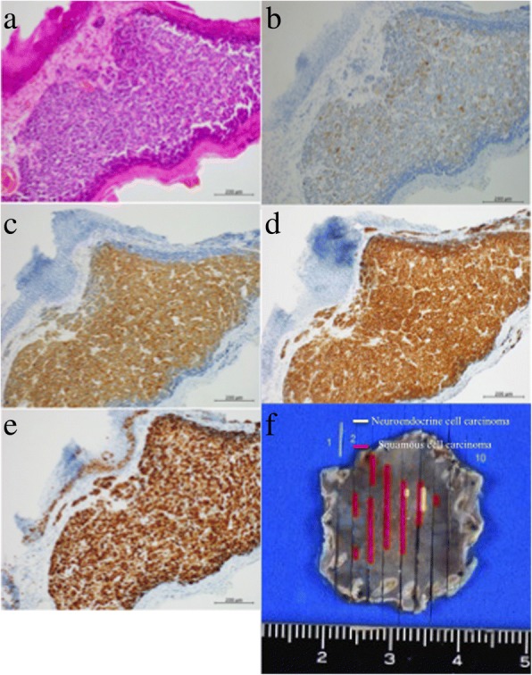
Fig. 2.

Resected specimen by endoscopic submucosal dissection. This specimen shows neuroendocrine cell carcinoma arranged in a sheet fashion with mixed squamous cell carcinoma. Neuroendocrine cell carcinoma formed a duct and it is surrounded by squamous cell carcinoma. a Hematoxylin and eosin staining. Immunohistochemical staining showing: b chromogranin A, c synaptophysin, d CD56, and e Ki-67. f The fixed resected specimen is mapped by yellow and red lines. Red lines squamous cell carcinoma, yellow lines neuroendocrine cell carcinoma
