Fig. 3.

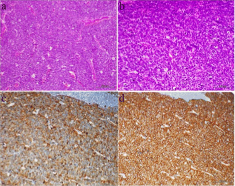
Histological examination of a resected lymph node. a Low-power view and b high-power view (hematoxylin and eosin staining). Immunohistochemical staining shows: b chromogranin A, c synaptophysin, and d CD56

Histological examination of a resected lymph node. a Low-power view and b high-power view (hematoxylin and eosin staining). Immunohistochemical staining shows: b chromogranin A, c synaptophysin, and d CD56