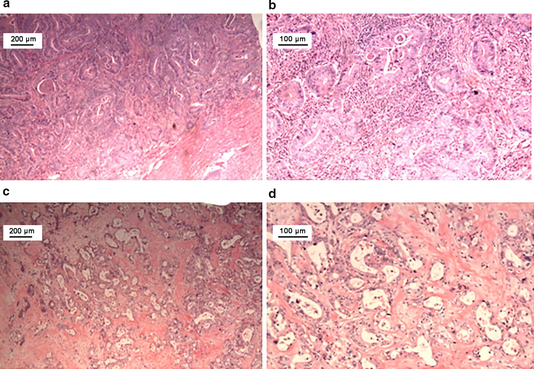
Fig. 2.

Histopathologic examination of the primary and metastatic duodenal adenocarcinoma by hematoxylin–eosin staining. a, b Microscopic examination reveals a moderately differentiated adenocarcinoma penetrating the visceral peritoneum and invading the pancreas. c, d Microscopic examination shows adenocarcinoma in the soft tissue of the superior mesenteric artery. a, c: ×40; b, d: ×100
