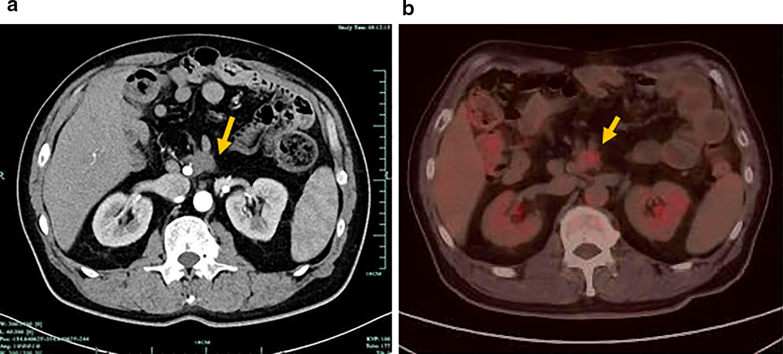
Fig. 3.

Computed tomography (CT) and PET/CT images of the metastatic lesion from a patient with duodenal adenocarcinoma. a CT scan shows the metastatic lesion (arrow) in the root of superior mesenteric artery and below transverse mesocolon. b PET/CT demonstrates malignant disease with FDG uptake (arrow) in the retroperitoneum
