
Figure 1.
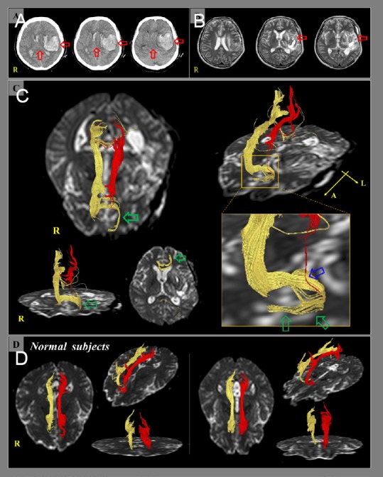
Brain computed tomography (CT), magnetic resonance (MR) imaging, and diffusion tensor tractography (DTT) of a 62-year-old female patient with a hemorrhagic stroke who showed a new neural tract between the injured anterior cingulums and the basal forebrain.
(A) Brain CT images at onset of hemorrhagic stroke show intracerebral hemorrhage in the left fronto-temporal lobe, intraventricular hemorrhage, subarachnoid hemorrhage, and subfalcine herniation (red arrow). (B) Brain MR images at three weeks after a hemorrhagic stroke show a leukomalactic lesion in the left fronto-temporal area (red arrow). (C) DTT of the cingulum. On 3-week DTT images, both cingulums show discontinuations between the anterior cingulums and the basal forebrain. However, the discontinued right anterior cingulum is connected to the left basal forebrain (green arrows) via the genu of the corpus callosum. In addition, the discontinued left anterior cingulum is connected to the unusual neural tract (blue arrow) from the right anterior cingulum connected to the left basal forebrain. (D) DTT of the fornix in two normal subjects (60- and 63-year-old females).