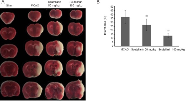
Figure 6.
Effects of scutellarin on infarct area of rats after cerebral ischemic injury.
(A) Representative images of TTC-stained brain sections (the infarcted region appears white). (B) Quantification of infarct area 3 days post-injury (% of contralateral hemisphere). Data are expressed as the mean ± SD (n = 11 per group; one-way analysis of variance followed by the Tukey-Kramer multiple comparison test). ##P < 0.01, vs. MCAO group. TTC: 2,3,5-Triphenyltetrazolium chloride; MCAO: middle cerebral artery occlusion.

