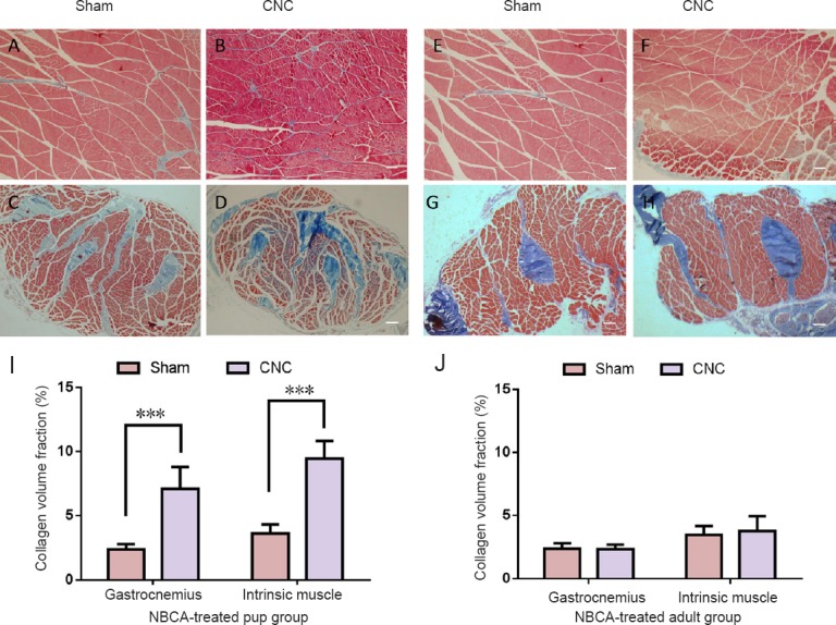
Figure 8.

Light microscopy analysis of the gastrocnemius and intrinsic muscles.
Light microscopy images of transverse sectioned muscle samples from the NBCA-pup and NBCA-adult groups following Masson's trichrome staining at 20 weeks after surgery. (A, B) Gastrocnemius and (C, D) intrinsic muscles of the hindpaw in the NBCA-treated pup group (n = 5). (E, F) Gastrocnemius and (G, H) intrinsic muscles of the hindpaw in the NBCA-treated adult group (n = 5). Scale bars: 100 μm. (I, J) Quantitative analyses of collagen volume fraction in the muscles on the sham-operated side and on the chronic nerve compression (CNC) side are presented for the pup (I) and adult (J) groups. All data are represented as the mean ± SD (repeated measures one-way analysis of variance followed by Dunnett's post hoc test). ***P < 0.001. NBCA: N-butyl-cyanoacrylate.
