Figure 2.

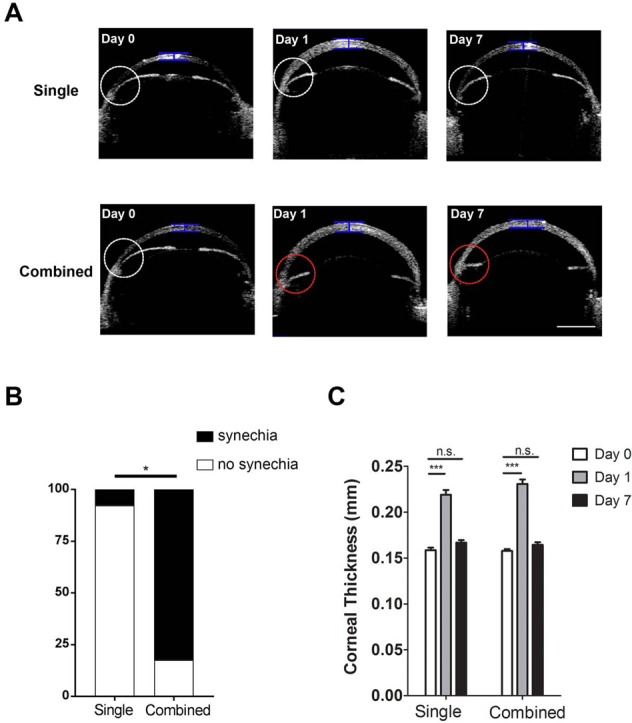
Anterior-segment OCT evaluation of central corneal thickness and anterior chamber angles in mice age 5 weeks. (A) Representative OCT images showing central corneal thickness and anterior chamber angles pre- (Day 0) and post-laser treatment on days 1 and 7 after the single or combined procedure. White circle indicates normal anterior chamber angle; red circle indicates synechiae at the angle with iris flattening; blue bar indicates site of measurement for central corneal thickness. Scale bar: 1 mm. (B) Summarized data showing significantly lower rate of synechiae in single than combined treatment. *P < 0.05 (n = 13–17/group). (C) Summarized and comparative data showing increased central corneal thickness at day 1 after single or combined treatment. No significant difference was found at day 7 after the procedures. ***P < 0.001. n.s., not significant (n = 10–12/group).
