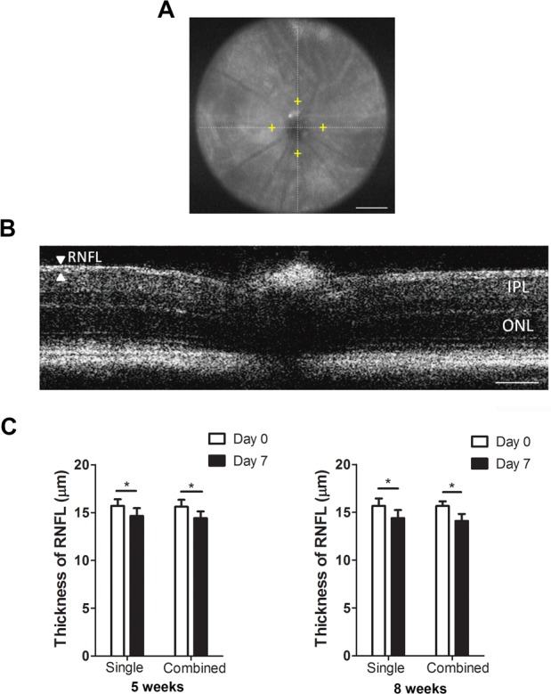
Figure 4.

Posterior-segment OCT evaluation on the thickness of RNFL after single or combined laser treatment in mice aged 5 or 8 weeks. (A) Representative OCT image showing a normal retina of a CD1 mouse. Yellow cross shows the measurement location at 400-μm distance from the optic nerve head. Scale bar: 500 μm. (B) Representative posterior-segment OCT image of the RNFL layer and total retina in a normal CD1 mouse. Scale bar: 100 μm. (C) Summarized data showing significant reduced RNFL thickness at day 7 post procedure in mice aged 5 or 8 weeks in both single and combined treatment settings. *P < 0.05 (n = 16–18/group). IPL, inner plexiform layer; ONL, outer nuclear layer.
