
Figure 2.
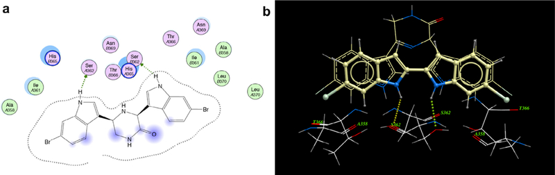
Binding mode of compound 4 at the MRSA PK tetramer interface binding site; (a) a two-dimensional map (MOE software) of the binding interactions between 1 and the interface site based on its co-crystallization with MRSA PK. Green arrows depict hydrogen-accepting interactions between 4 and MRSA PK residues from the interface (left). (b) Modeled overlay of compound 10b with 4 in the MRSA binding site (right) (ICM software from PDB data file 3t07).