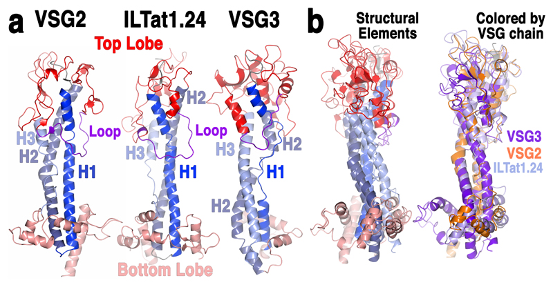
Fig. 1. Substantial structural divergence between VSGs.
(a) Comparison of monomeric VSG structures. VSG2 (MITat1.2), ILTat1.24 and VSG3 are shown as ribbon diagrams colored by six structurally conserved elements: the three helices of the stalk-like bundle (three shades of blue for each individual helix, H1-H3), the top and bottom lobes (red and salmon, respectively), and a conserved, elongated loop in the top lobe (purple). (b) Superposition of the three monomers, colored on the left by the schema described in (a) and on the right by individual protein as indicated in the labeling.

