
Figure 1.
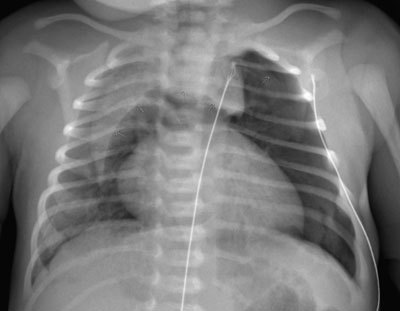
Frontal plain radiograph of the chest, showing the Spinnaker-Sail Sign. Both lobes of the thymus are lifted and displaced into the upper mediastinum, creating a wedge-shaped opacity. This resembles a Spinnaker sail. A pneumothorax can be seen at the left lung apex.