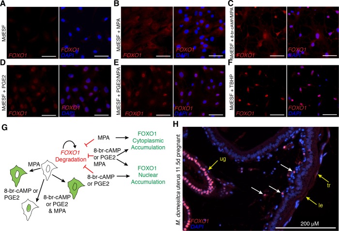
Fig 5. Post-translational regulatory control of FOXO1 by PGE2 and oxidative stress in MdESF.
(A-H) Immunofluorescence of FOXO1 in MdESF and pregnant M. dometica uterus. (A) FOXO1 is not detected above background in unstimulated MdESF. (B) FOXO1 protein is detected in the cytoplasm but not in the nucleus in MdESF treated for 2 days with MPA alone. (C) FOXO1 translocates to the nucleus and is detected in the cytoplasm in MdESF treated with decidualzing stimuli 8-br-cAMP/MPA for 2 days. (D-E) FOXO1 is detected in the nucleus and cytoplasm in MdESF treated for 2 days with either PGE2 alone or PGE2/MPA. (F) Oxidative stress induces FOXO1 to translocate to the nucleus in MdESF treated with tert-butyl hydrogen peroxide TBHP for 2 hours. Scale bars are 10 μm. (G) Model showing post-translational regulatory control of FOXO1 in MdESF treated for 2 days with stimuli in this study. (H) FOXO1 immunofluorescence of uterine tissue in cross section during late gestation (11.5 d.p.c.). All samples counterstained with DAPI. Arrows indicate FOXO1 detection. cAMP, cyclic AMP; d.p.c., days post coitus; FOXO, forkhead box class O; le, luminal epithelium; MPA, medroxyprogesterone acetate; PGE2, prostaglandin E2; ug, uterine glands; TBHP, tert-butyl hydrogen peroxide; tr, trophoblast.

