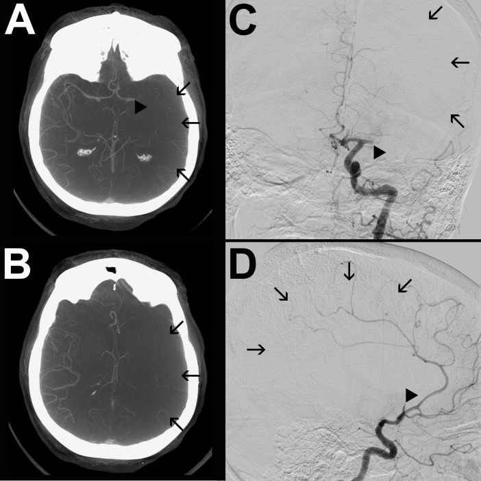
Fig 2.
mpFDCTA (A, B) and DSA images in posterior-anterior (C) and lateral projection (D) of a patient with poor collaterals are shown. The arrowheads (A, C) indicate an occluded M1-segment of the left MCA. There are no collaterals identifiable in the ischemic hemisphere. The arrows (A-D) indicate the ischemic bed with missing collateral vessels. Thus, the patient was assigned a collateral score of 0 on both, mpFDCTA and ASITN DSA score.

