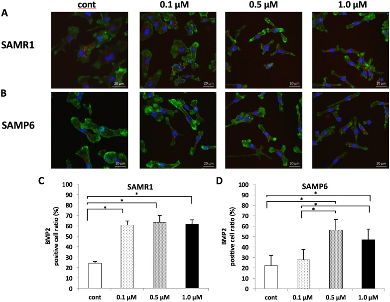
Fig 2. The protein expression of BMP2 detected with immunofluorescence staining (A, B) and percentages of BMP2 positive cells for 3days fluvastatin treatment in immunofluorescence staining (C, D).
(A, B) BMP2 expressions were found in the cytoplasm. Red: BMP2, Blue: DAPI and green: actin. Scale bar = 20 μm. (C, D) The percentage of BMP2-positive stained cells was significantly higher at concentrations more than 0.1 μM in SAMR1 and at concentrations more than 0.5 μM in SAMP6. *P < 0.05.

