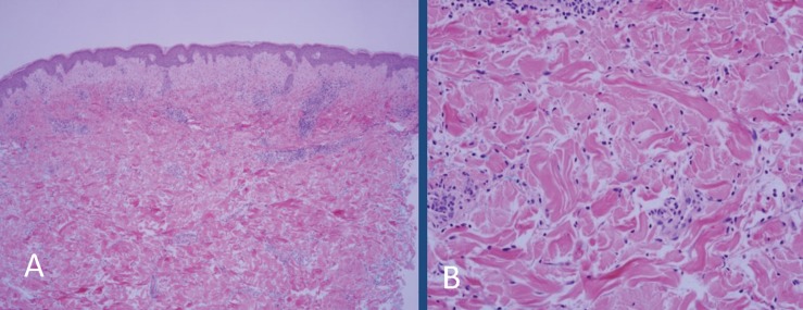
Figure 2.
(A) Biopsy of the left lateral abdomen shows mild superficial dermal edema and interstitial and perivascular inflammation (10X, hematoxylin and eosin). (B) High power image showing that the majority of the inflammatory cells are interstitial and perivascular neutrophils. The blood vessels fail to show vascular damage (20X, hematoxylin and eosin).

