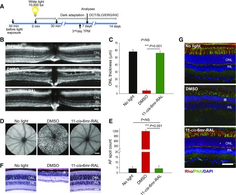
Fig. 1.
Protective effect of 11-cis-6mr-retinal against bright-light–induced retinal degeneration in Abca4−/−Rdh8−/− mice. (A) Scheme of mouse treatment. 11-cis-6mr-retinal or DMSO vehicle was administered to 4–6-week-old Abca4−/−Rdh8−/− or WT BALB/c mice by intraperitoneal injection 30 minutes before exposure to bright light (10,000 lux) for 30 minutes for Abca4−/−Rdh8−/− mice and 12,000 lux for 60 minutes for WT BALB/c mice. Then, mice were kept in the dark for 7–14 days before examination. The morphology of the retina was assessed by OCT and SLO in vivo imaging, as well as by H&E staining and immunohistochemistry. Retinal function was assessed by ERG. TPM imaging was used to determine abnormalities in photoreceptor cells on the 4th day after treatment. (B) Representative OCT images obtained 7 days after treatment of mice with 11-cis-6mr-retinal (20 mg/kg b.wt.) 30 minutes before exposure to white light at 10,000 lux for 30 minutes. After light illumination, mice were kept in the dark until morphologic examination. INL, inner nuclear layer. Asterisk indicates severely disrupted photoreceptor structures in DMSO-treated control mice. (C) Quantification of the ONL thickness obtained in 10 mice per treatment group. Error bars indicate standard deviations. Changes in the ONL thickness observed after treatment with 11-cis-6mr-retinal compared with DMSO-treated group were statistically significant (P < 0.001); no significant difference in the ONL thickness was observed between animals unexposed to light and those treated with 11-cis-6mr-retinal (P = NS). Statistical significance was calculated with the one-way analysis of variance and post hoc Student’s t test, unpaired two-tailed. (D) Representative SLO images show AF spots in the retina of a mouse exposed to light after pretreatment with DMSO (center). Mice unexposed to bright light (left) or exposed to bright light after pretreatment with 11-cis-6mr-retinal (right) exhibited much fewer spots. (E) Quantification of AF spots performed in 10 mice per treatment group. Error bars indicate standard deviation. Changes in the number of AF spots after treatment with 11-cis-6mr-retinal compared with the DMSO-treated group were statistically significant (P < 0.001). No significant difference was observed between animals unexposed to light and those exposed to light after treatment with 11-cis-6mr-retinal (P = NS). Statistical significance was calculated with one-way ANOVA and post hoc, the Student’s t test, unpaired two-tailed. (F) Examination of retinal morphology after staining with H&E in paraffin sections prepared from eyes collected from Abca4−/−Rdh8−/− mice either unexposed to light or exposed to bright light after the indicated treatments. Asterisk indicates severely disrupted photoreceptor structures in DMSO-treated control mice. (G) Examination of retinal morphology by immunohistochemistry in cryosections prepared from the eyes collected from Abca4−/−Rdh8−/− mice either unexposed to light or exposed to bright light after the indicated treatments. Sections were stained with an anti-rhodopsin C terminus specific antibody (red), which indicates the structural organization of rod photoreceptors; PNA staining (green), which indicates the health of cone photoreceptors; and DAPI staining of nuclei (blue). Asterisk indicates severely disrupted photoreceptor structures in DMSO-treated control mice. Scale bar, 50 µm.

