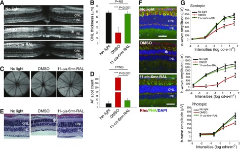
Fig. 4.
11-cis-6mr-retinal prevents bright light-induced retinal degeneration in WT mice. BALB/c mice were injected intraperitoneally with 11-cis-6mr-retinal (20 mg/kg b.wt.) 30 minutes before exposure to white light at 12,000 lux for 1 hour and then kept in the dark for 7 days before examination of retinal morphology and function. (A) Representative OCT images show substantial protection of the ONL in mice pretreated with 11-cis-6mr-retinal compared with DMSO-treated control mice. Asterisk indicates severely disrupted photoreceptor structures in DMSO-treated control mice. Scale bar, 50 µm. (B) Quantification of the ONL thickness in mice unexposed to light or in mice treated with either DMSO vehicle or 11-cis-6mr-retinal before exposure to bright light in five mice per each treatment group. Error bars indicate standard deviation. Statistical significance was calculated with one-way ANOVA and post hoc Student’s t test, unpaired two-tailed. (C) Representative SLO images show AF spots in the retina of mice unexposed to light or exposed to bright light after pretreatment either with DMSO vehicle or 11-cis-6mr-retinal. Scale bar, 50 µm. (D) Quantification of AF spots in five mice per treatment group. Error bars indicate standard deviations. Changes in the number of AF spots after treatment with 11-cis-6mr-retinal compared with DMSO-treated group were statistically significant (P < 0.001). No significant difference was observed between mice unexposed to light and those treated with 11-cis-6mr-retinal (P = NS). Statistical significance was calculated with one-way ANOVA and post hoc Student’s t test, unpaired two-tailed. (E) Examination of gross retinal morphology after H&E staining of paraffin sections from eyes collected from WT mice either unexposed to light or exposed to bright light after indicated treatments. Asterisk indicates severely disrupted photoreceptor structures in DMSO-treated control mice. Scale bar, 50 µm. (F) Examination of retinal morphology by IHC in cryosections prepared from eyes of BALB/c mice either unexposed to light or exposed to bright light after indicated treatments. Sections were stained with an anti-rhodopsin C terminus specific antibody (red) that indicates the structural organization of rod photoreceptors. PNA staining (green) denotes the health of cone photoreceptors, and DAPI staining (blue) reveals the nuclei. Asterisk indicates severely disrupted photoreceptor structures in DMSO-treated control mice. Scale bar, 50 µm. (G) Visual function determined by ERG responses. ERG recordings revealed significant protective effects in 11-cis-6mr-retinal-treated mice before acute-light illumination as compared with DMSO-treated mice in both scotopic a- and b-waves and in photopic b-waves. ERG measurements were carried out in five mice per group.

