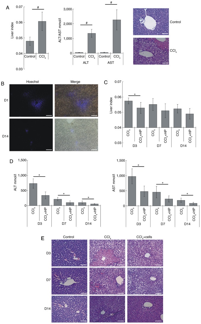
Figure 7.
HP14-19-CD cell transplantation improves liver functional recovery in mice with acute liver injury. (A) Acute liver failure model was established by using 2% CCl4 gavage. After 24 h, the liver index (liver wet weight/body weight ×100%), serum levels of ALT and AST, and the pathological changes in liver were determined to evaluate liver structure and function. Scale bar, 200 µm. (B) Hoechst 33342-pre-labelled cells were injected into the splenic vein. The survival and distribution of exogenous cells were detected on day 1 and at 14 days after transplantation. Scale bar, 200 µm. (C) The liver index of each group was determined at the indicated time points. (D) The serum levels ALT and AST in each group were detected at the indicated time points. (E) The specimens were fixed in 4% paraformaldehyde, embedded in paraffin following dehydration and serially cut into 5-µm-thick sections. Serial sections were stained with hematoxylin and eosin. Scale bar, 200 µm. #P<0.05 vs. control group; *P<0.05 vs. CCl4 model group. ALT, alanine aminotransferase; AST, aspartate amino-transferase; CD, cytosine deaminase; D, day.

