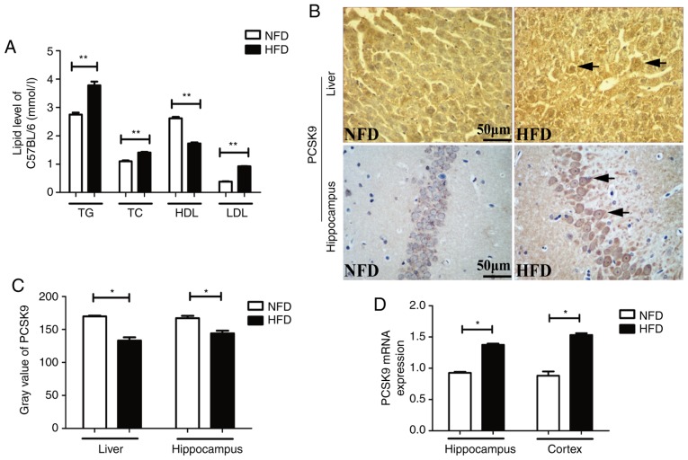
Figure 1.
Blood lipids and PCSK9 expression levels were significantly increased upon HFD feeding. (A) Serum TG, TC and LDL-C levels were significantly increased, while HDL-C level was significantly decreased, in HFD mice compared with those in NFD mice (n=8). (B) Representative PCSK9 IHC staining in the liver and hippocampus tissues following HFD feeding. (C) Gray value of PCSK9 in IHC analysis was significantly decreased in the liver and hippocampus upon HFD feeding (n=4). (D) PCSK9 mRNA expression was significantly increased in the hippocampus and cortex following HFD feeding, according to reverse transcription-quantitative polymerase chain reaction analysis (n=4). *P<0.05 and **P<0.01 vs. NFD mice. PCSK9, proprotein convertase subtilisin/kexin type 9; HFD, high-fat diet; NFD, no-fat diet; TG, triglycerides; TC, total cholesterol; LDL-C, low-density lipoprotein cholesterol; HDL-C, high-density lipoprotein cholesterol; IHC, immunohistochemistry.

