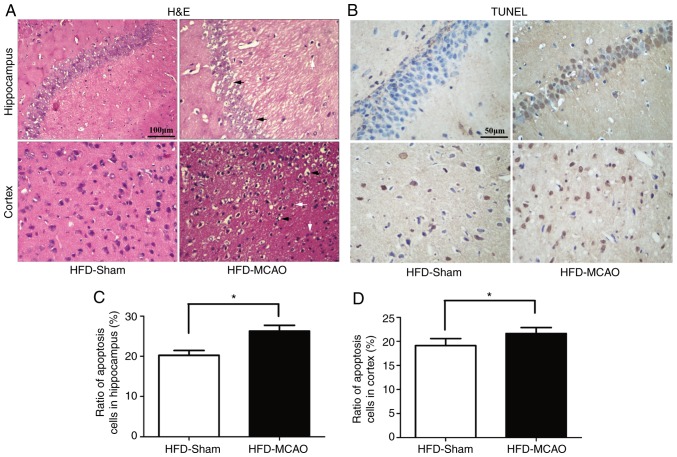
Figure 3.
Ischemic injury and increasing apoptosis were observed in the hippocampus and cortex following middle cerebral artery occlusion in hyperlipidemic mice. (A) Representative images of the pyknotic cells and neuronal vacuolization in the hippocampus and cortex. (B) Representative images of the TUNEL-positive nuclei (brown) and total nuclei (blue). Bar plots demonstrating the percentage of TUNEL staining in the (C) hippocampus and (D) cortex. Black arrows indicate neuronal vacuolization and white arrows indicate inflammatory infiltration. *P<0.05 and **P<0.01 vs. HDF-sham mice (n=4). TUNEL, terminal deoxynucleotidyl transferase dUTP nick end labeling; HFD, high-fat diet.

