
Fig. 1. AOM-injected mice developed FHF.
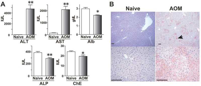
(A) Blood levels of ALT, AST, Alb, ALP and ChE at 24 h after AOM injection are shown (Control naive mice, n = 6; AOM-treated mice, n = 4). Mean + SEM, *, P< 0.05, **, P < 0.01 versus control mice by Mann-Whitney U test. (B) Histopathological changes of liver were observed 24 h after AOM exposure. Hematoxylin and eosin staining shows the severe hemorrhage (arrows), microvesicular steatosis and centrilobular necrosis in AOM-treated mice (Bars, 100 μm).