
Fig. 3. LPA disrupted BBB barrier function in in vitro BBB model.
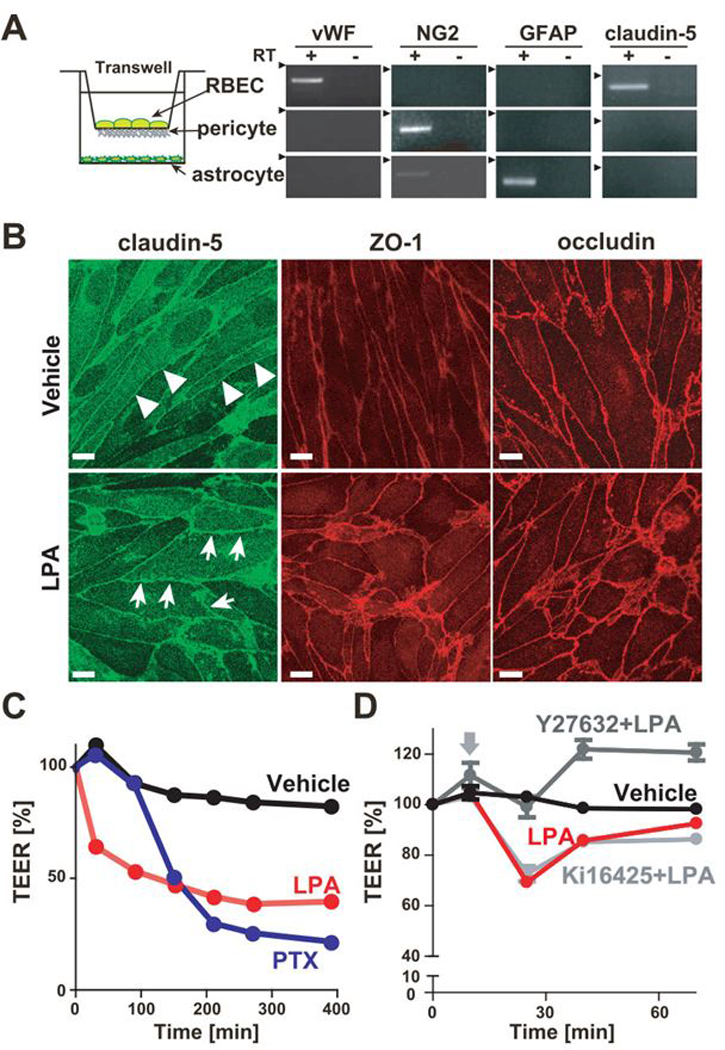
(A) Schematic representation of the in vitro BBB model is shown. mRNA expression in three primary cultured cells were examined by RT-PCR. (B) Immunofluorescent staining of confluent RBECs in BBB model for tight junction proteins ZO-1, claudin-5 and occludin. Cells were immunostained 15 min after 50 μM LPA stimulation. Arrows show zipper-like staining between LPA-treated RBECs, while the junctional immunostaining of RBECs forms a continuous, smooth and belt-like pattern (arrowheads) in vehicle-treated RBECs (Bar, 10 μm). (C) Change of TEER in BBB model treated with 50 μM LPA and 200 ng/ml PTX was evaluated by an epithelial-volt-ohm meter. Data is representative of three independent experiments with similar results. (D) Change of TEER in BBB model was evaluated by an epithelial-volt-ohm meter. Cells were pretreated with or without 10 μM Y-27632 and 10 μM Ki16425 for 10 min and then stimulated with 50 μM. Data is representative of three independent experiments with similar results.