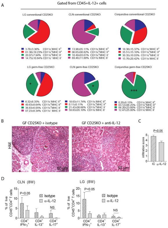
Figure 5. IL-12 Neutralization in Germ-free CD25KO mice Improves Autoimmunity.
A-Single cell suspensions were prepared from conventional and germ-free lacrimal glands (LG), cervical lymph nodes (CLN) or conjunctiva and stained with live/dead dye and CD45, IL-12, CD11c, and MHC II antibodies. Single cells, alive CD45+IL-12+ cells were gated and CD11c and MHC II were plotted.
B–D-Germ-free CD25KO mice, aged four weeks of age, received either anti-IL-12 (α-IL-12) or isotype control (IC) antibody bi-weekly i.p. injections (200ug/injection) for a total of 4 weeks while kept in a germ-free isolator. Mice were used at eight weeks of age (n = six/group).
(A) Flow cytometry analysis showing distribution of CD45+IL-12+ cells based on CD11c and MHC II expression; pie charts of means of four samples. Means and SD are shown below. Nonparametric Mann–Whitney U tests were used to make comparisons between germ-free and conventional mice, * P<0.05, ***P<0.001.
(B) Representative pictures of haematoxylin and eosin (H&E)-stained sections of the lacrimal gland. 20X objective, scale bar = 50 μm. (C) Inflammation scores of lacrimal gland pathology. Bar graphs show means ± SEM of two independent experiments, final n = five animals. Nonparametric Mann–Whitney U tests were used to make comparisons.
(D) Flow cytometric analysis of cervical lymph nodes and lacrimal gland. Bar graphs are means ± SD of two independent experiments, with a final sample size of six per group. Parametric t-tests were used to make comparisons between groups.
LG = lacrimal gland; CLN = cervical lymph nodes; NS = non-significant

