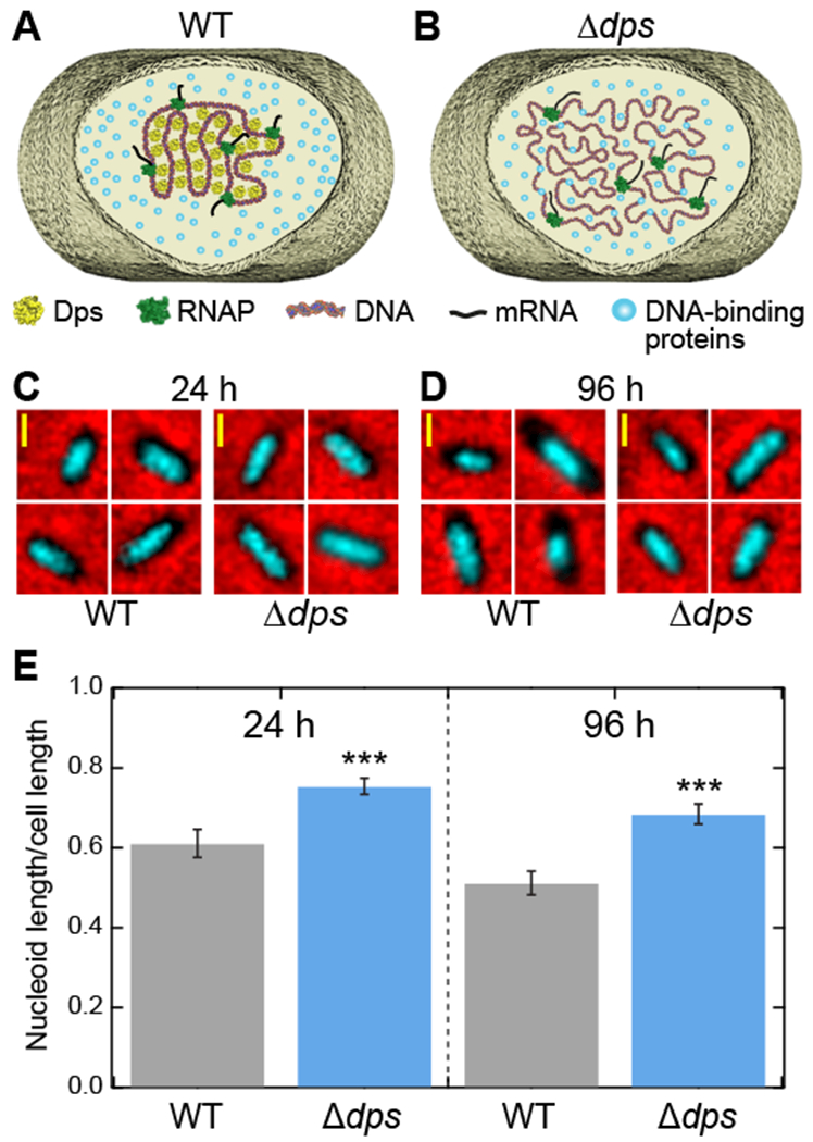
Figure 1. Dps compacts the nucleoid in stationary-phase E. coli.

(A) Schematic of the structure of DNA in a wild-type cell during stationary phase. Dps condenses the cellular DNA. (B) In Δdps cells, Dps-mediated DNA compaction cannot occur. (C, D) Fluorescence images of the nucleoid from wild-type and Δdps cells stained with Hoechst 33258 (cyan) were superimposed onto phase-contrast images of the same cells (black on red) grown for (C) 24 hours or (D) 96 hours. (E) Ratios of nucleoid length to cell length, extracted from fluorescence images (n = 133 - 208 cells per condition). The error bars represent the estimate of the standard errors by bootstrapping. See also Figure S1.
