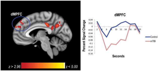
Figure 2.
Compared to controls, the mTBI group had significant deactivation in default mode network regions for the incorrect > correct contrast. In particular, the mTBI group had greater deactivation in the left dMPFC and left PCC. The hemodynamic response function for the dMPFC is plotted to the right of the figure. dMPFC=dorsomedial prefrontal cortex; mTBI=mild traumatic brain injury; PCC=posterior cingulate cortex.

