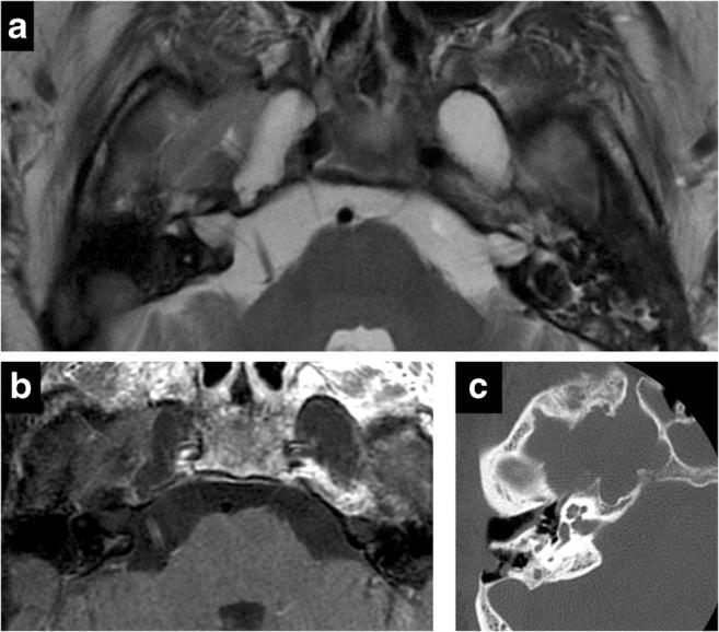
Fig. 10.

Dural ectasia in neurofibromatosis type 1. Axial T2 (a), axial C+ (b), CT (c). Bilateral enlargement of Meckel’s cave—CSF isointense and no abnormal enhancement. Smooth scalloping and remodelling of petrous apex on CT (c)

Dural ectasia in neurofibromatosis type 1. Axial T2 (a), axial C+ (b), CT (c). Bilateral enlargement of Meckel’s cave—CSF isointense and no abnormal enhancement. Smooth scalloping and remodelling of petrous apex on CT (c)