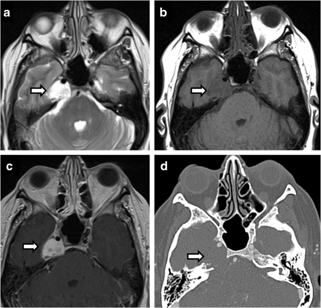
Fig. 19.
Chondrosarcoma. Axial T2 (a), axial T1 (b), axial T1 C+ (c), CT (d). A 61-year-old woman with effacement of the right Meckel’s cave by an expansile petrous apex mass that is hyperintense on T2, hypointense on T1 and shows avid enhancement on post contrast image. CT shows features of slow growing lesion. Note preserved Meckel’s cave on the left

