
Fig. 2.
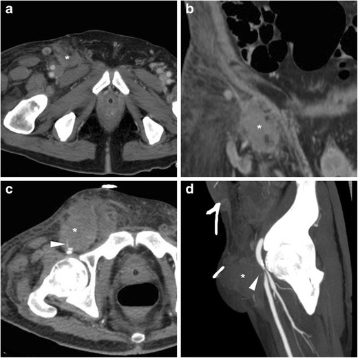
Delayed groin bleeding in a 70-year-old male patient after transarterial mitral and aortic valve replacement. Early CT (A, B) showed circumscribed effusion (*) abutting the CFA at the level of percutaneous access, which was interpreted as a minor bleed and treated conservatively. However, during hospitalisation for infected sternal dehiscence, repeated CT angiography (C, D) showed development of a 4.5-cm groin haematoma (*) containing a thin contrast medium (CM) extravasation focus (arrowheads), causing compression on the CFA (MIP image D) which required stenting