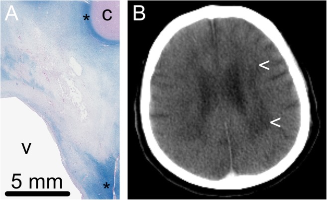
Fig. 4.

Cerebral white matter lesions. a Histological slides (Luxol fast blue-Pas stain) showed dispersion of the matrix with extensive demyelination in the central white matter. Normal myelination is seen subcortically and in the area around the basal nuclei (*). c = cortex, v = ventricle. b The CT scan of the patient showed nonspecific white matter abnormalities (<)
