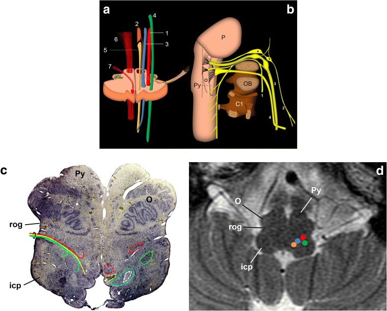
Fig. 1.
Schematic representation of the glossopharyngeal nerve. a Transversal section of the medulla oblongata at the level of the inferior olivary nucleus. Efferent nuclei: nucleus ambiguous (1) and inferior salivatory nucleus (2); afferent nuclei: solitary nucleus (3) and sensitive trigeminal nucleus (4). Other relevant structures: dorsal motor nucleus of the vagus nerve (5), pyramidal tract (6) and hypoglossal nerve (7). b Schematic drawing of the lower cranial nerves representing their mutual relationships and course from the brain stem to the cranial exit. 1: spinal nerve; 2: glossopharyngeal nerve; 3: vagus nerve; 4: hypoglossal nerve; P: pons; O: inferior olivary nucleus; Py: pyramid; C1: atlas; OB: occipital bone. The hypoglossal nerve exits the cranium through the anterior condylar canal; the glossopharyngeal, vagus and spinal nerves exit the cranium along the jugular foramen. *Glossopharyngeal nerve nodes; black dotted line: retro-olivary groove where the glossopharyngeal nerve exits the medulla oblongata with the vagus and spinal nerves. The white dotted line draws the groove between the pyramid and the inferior olivary nucleus, where the hypoglossal nerve exits the brain stem. c, d Upper medulla oblongata as viewed from below. Micrographic slice showing the glossopharyngeal nerve nuclei (c) and axial T2-weighted brain MRI at the same level (d). In the anatomical slice (Nissl stain), left nuclei are circumscribed by dotted lines. All nuclei are behind the retro-olivary groove (rog). The nerve’s pathway is represented on the right side of the sample, between the retro-olivary nucleus and the inferior cerebellar peduncle (icp). Colours as in a (the white dotted circle encloses the solitary tract)

