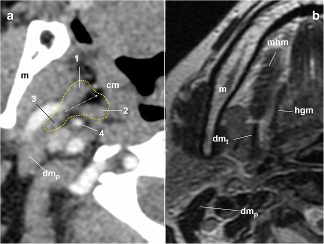
Fig. 10.
Cervical portion of the glossopharyngeal nerve. a Axial contrast-enhanced CT scan of the neck, right side, soft tissues window. The dotted yellow line draws the limits of the styloid pyramid. 1: styloglossus muscle; 2: stylopharyngeus muscle; 3: stylohyoid muscle; 4: external carotid artery; cm: constrictor muscles. The dotted white arrow represents the long axis of the styloid pyramid as a reference of the gpn pathway. dmp: posterior belly of the digastric muscle; m: mandible. b Axial T2-weighted MRI of the neck, right side, at the level of the intermediate tendon of the digastric muscle (dmt). The slice over the level of the tendon is a good reference for the entry point of the glossopharyngeal nerve in the pharynx. mhm: mylohyoid muscle

