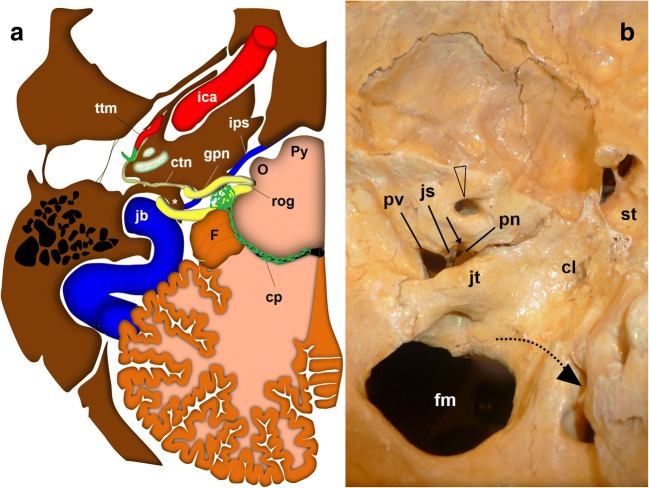
Fig. 2.
The glossopharyngeal nerve (gpn), cisternal portion. a Schematic drawing of a transversal slice at the level of the medulla oblongata (view from above). The gpn exits the medulla at the retro-olivary groove (rog) and crosses through the cerebellomedullary cistern to the jugular foramen. Within the cistern, it lays below and in front of the choroid plexus (cp) of the fourth ventricle and the cerebellar flocculus (F). The glossopharyngeal nerve exits the skull medial and anterior to the jugular spine (*). At that level, it is closely related to the inferior petrosal sinus (ips). The glossopharyngeal nerve crosses first over the sinus to be anterior to the sinus at the extracranial verge of the jugular foramen, posterior to the internal carotid artery. ctn: caroticotympanic or Jacobson nerve; ica: internal carotid artery; jb: jugular bulb; mo: medulla oblongata; O: inferior olivary nucleus; P: Pons; Py: pyramid; ttm: tensor tympani muscle; va: vertebral arteries. b Skull base, view from inside. The lower cranial nerves have a close relationship with the jugular tubercle (jt). The glossopharyngeal nerve (represented by the dotted black arrow) exits through the pars nervosa of the jugular foramen, split from the pars vascularis (pv) by the jugular spine (js). cl: clivus; fm: foramen magnum; st: sella turcica; arrowheads: internal auditory meatus

