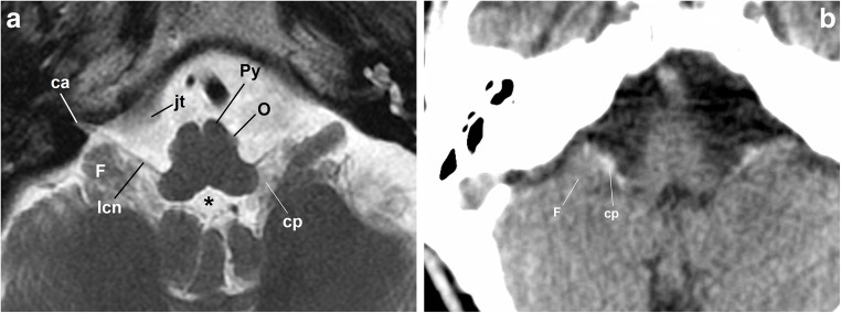
Fig. 3.
a Axial fast spin-echo T2-weighted brain MRI at the level of the medulla oblongata. The image shows the lower cranial nerves (lcn) passing by the flocculus (F), the choroid plexus (cp) outside the fourth ventricle (*) and the jugular tubercle (jt) as they approach the cochlear aqueduct (ca); O: inferior olivary nucleus; Py: pyramid. b Axial non-enhanced brain CT scan, brain window. The flocculus (F) and the choroid plexus (cp) are reliable references to determine the position of the nearby glossopharyngeal nerve

